Healthy Soch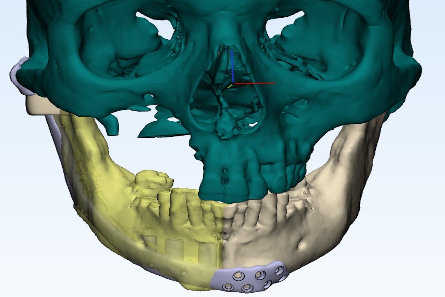
After extensive surgery and radiotherapy for Stage III oral cancer, a 49-year-old man was left with a severe mandibulofacial defect, persistent oronasal fistula, and restricted mouth opening
Using 3D virtual surgical planning, a customized titanium implant and a free anterolateral thigh flap, doctors performed a complex secondary reconstruction aimed at restoring his facial structure, speech, swallowing, mastication and quality of life
India
healthysoch
Mumbai/New Delhi, October 08, 2026:
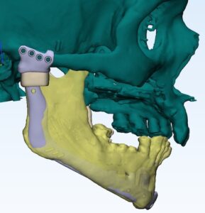 A complex facial deformity following cancer treatment left a 49-year-old cancer survivor struggling with essential functions such as eating, speaking and swallowing, making reconstruction critical to restoring both his appearance and quality of life. A multidisciplinary team led by Dr. Amit Chakraborty, Lead Head & Neck Surgical Oncologist at SSO Cancer Hospitals, Performed a complex secondary jaw reconstruction using 3D virtual surgical planning, a customized patient-specific titanium implant, and a left free anterolateral thigh flap, helping restore the patient’s facial structure and functional abilities.
Patient Mr. Arvind Tagare, a 49-year-old retired engineer from Indore, was diagnosed with Stage III carcinoma of the right buckle mucosa (inner lining of the right cheek) in March 2024. He underwent extensive cancer surgery followed by multimodality treatment. He later developed a postoperative oronasal fistula (an abnormal opening between the mouth and nose) with nasal regurgitation (food or liquids passing into the nose), along with a major defect involving the jaw and face. The condition affected his ability to eat, speak, and swallow and impacted his facial appearance and quality of life.
Dr. Amit Chakraborty, Lead Head & Neck Surgical Oncologist at SSO Cancer Hospitals, Mumbai Said, “A PET-CT confirmed his current disease-free status and ruled out recurrence or metastasis, allowing the medical team to focus on complex reconstructive rehabilitation. Without reconstruction, the persistent oronasal fistula could have resulted in worsening nasal regurgitation, compromised mastication and swallowing, nutritional deterioration, progressive facial disfigurement and a substantial decline in quality of life. The reconstructive objective was therefore not simply to replace the missing jaw, but to restore facial architecture, symmetry, oral competence, mastication, speech, swallowing and appearance, helping the patient regain functional independence and confidence.”
The reconstruction was particularly challenging as it involved a previously operated and irradiated field, with severe radiation fibrosis, distorted tissue planes, restricted mouth opening and potentially compromised recipient vessels. To overcome these challenges, the surgical team used 3D virtual surgical planning to visualize the exact defect, assess facial asymmetry and determine the ideal reconstructive contour before surgery. Using the patient’s CT-derived DICOM dataset, a customized biocompatible titanium patient-specific implant was digitally designed to recreate his individual mandibulofacial anatomy. A left free anterolateral thigh flap was used to provide vascularized soft-tissue coverage, with the customized implant prepared four days before surgery.
Dr Amit Added, “This was an exceptionally demanding secondary reconstruction because the patient had previously undergone oncological surgery and radiotherapy, leaving severe fibrosis, scarred and distorted tissue planes, restricted mouth opening and potentially compromised recipient vessels. The objective was not merely to replace the missing portion of the jaw, but to restore the mandibular framework, facial contour, symmetry, and soft-tissue volume, while improving oral competence, mastication, speech, and swallowing. In such complex secondary defects, conventional reconstruction can be challenging because it relies more heavily on intraoperative estimation and manual bending. Three-dimensional virtual surgical planning allowed us to analyses the patient’s native craniofacial anatomy and contralateral symmetry before surgery and digitally recreate the desired facial projection and contour. A customized titanium patient-specific implant was then designed from his CT data to achieve greater anatomical accuracy, predictability and reproducibility during reconstruction.”
Dr Amit further added, “The patient-specific titanium implant was combined with a left free anterolateral thigh flap to provide vascularized soft-tissue coverage. The procedure was completed uneventfully, although the reconstruction required meticulous planning because of the previous radiation, fibrosis, altered tissue planes, limited mouth opening and changes in the recipient vessels. Postoperatively, Ryle’s tube feeding was continued for approximately two weeks to protect the intraoral reconstruction, minimize stress on the suture line and support optimal tissue healing. The patient was discharged 30th August 2026. and is now undergoing structured follow-up at one week, two weeks, one month and thereafter under oncological and reconstructive surveillance. Rehabilitation will focus on progressively improving mastication, speech and swallowing and helping him return towards functional independence and normal daily activities.”
“In modern head and neck cancer care, removing the cancer is only the first step. True recovery also means helping the patient regain essential functions such as eating, speaking and swallowing, while restoring facial appearance, confidence and independence. In this case, the use of 3D virtual planning, a customized titanium implant and microsurgical reconstruction allowed us to rebuild the jaw and facial structure with greater precision, helping the patient move towards a better quality of life and return to everyday activities with dignity,” concluded Dr Amit Chakraborty.