19kg Ovarian Tumor Removed in Rare, High-Risk Surgery at HCG Panda Cancer Hospital
India
healthysoch
Cuttack/New Delhi, June 27, 2025:
 In a landmark of surgical oncology achievement, the multidisciplinary oncology team at HCG Panda Cancer Hospital, Cuttack, successfully removed a massive 19-kilogram ovarian tumor from a 54-year-old woman with significant cardiac comorbidities, saving her life and significantly improving her quality of health.
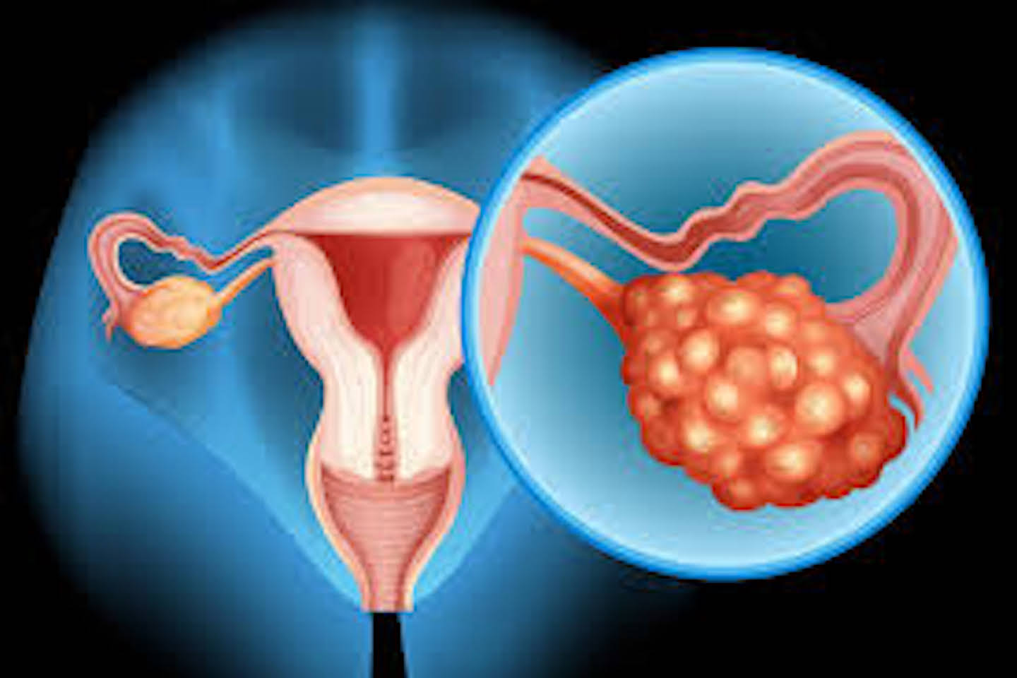
The patient had been experiencing symptoms such as abdominal bloating and discomfort for nearly five months before seeking medical attention. Her first visit to HCG Panda Cancer Hospital was in December 2024. Prior to presenting at HCG, the patient had undergone initial evaluation at other healthcare facilities, including FNAC, PET-CT, and tumor marker testing.
The patient had presented with progressive abdominal distension, persistent pain, and shortness of breath—symptoms that had been worsening for months and severely impacting her day-to-day life. Diagnostic imaging revealed a large ovarian mass occupying nearly the entire abdominal cavity, displacing vital organs and exerting pressure on the diaphragm and gastrointestinal tract.
PET-CT imaging was the primary modality used to assess tumor spread. A guided FNAC revealed metastatic adenocarcinoma deposits, and tumor marker levels were elevated—CEA (Carcinoembryonic Antigen): 40.2, CA-125: 352—confirming a malignant ovarian tumor with serosal and omental deposits, including in the pouch of Douglas.
Initially, she was administered systemic chemotherapy and targeted therapy in an effort to shrink the tumor. However, her disease showed no response to medical treatment, leading to considerable emotional and physical distress for the patient and her family. With the tumor continuing to grow and medical options exhausted, surgery became the only curative path—albeit a high-risk one.
“The surgical decision was complex and high stakes,” said Dr. Jyotiranjan Swain, senior surgical oncologist at HCG Panda Cancer Hospital and lead surgeon for the case. “The tumor was not only massive in size but also posed a direct threat to multiple organ systems. To make matters more critical, the patient was a known case of paroxysmal supraventricular tachycardia (PSVT)—a cardiac arrhythmia that significantly increased intraoperative risk.”
Adding to the complexity was the fact that the tumor had shown poor response to neoadjuvant chemotherapy, including bevacizumab, which carries a known risk of causing intestinal perforation during surgery. Furthermore, the size of the mass caused such severe compression that the patient could not even change posture comfortably.
The five-hour-long procedure was meticulously planned and executed by a dedicated team comprising surgical, anesthesia, and critical care experts. The anesthesia team, led by Dr. Sandeep Kumar Panda, played a pivotal role in maintaining hemodynamic stability and respiratory function throughout the procedure. Given the size of the mass and its proximity to the diaphragm, the risk of intraoperative respiratory distress and arrhythmia was substantial.
“This was a highly delicate case from the anesthesia perspective,” noted Dr. Panda. “Preoperative optimization, careful cardiac monitoring, and real-time coordination with the surgical team were critical. We had to constantly anticipate fluctuations in heart rate and respiratory mechanics due to the tumor’s sheer bulk and anatomical location.”
The surgery was completed successfully with no major intraoperative complications, and the tumor—one of the largest ever operated on at the center—was fully excised, despite extensive intra-abdominal involvement.
Post-operative care was managed under the close supervision of Dr. Subhankar Das, Consultant Intensivist, and the critical care team, who ensured a stable recovery during the high-risk post-surgical period.
The patient’s total hospital stay was 22 days, with approximately 7 days spent on preoperative optimization. She was monitored in the ICU for 3 days post-surgery and was discharged on the 16th post-operative day. Mobilization began as early as the 2nd post-op day, and she resumed basic activities by the 10th day.
The patient made a remarkable recovery and was discharged with a positive prognosis. She continues to receive regular follow-up care and has shown significant improvement in overall health and mobility.
The patient is currently planned for adjuvant chemotherapy with a FOLFIRI regimen and will be followed up at three-month intervals, including physical examinations, tumor marker assessments, and imaging as needed.
Dr. Swain emphasized the importance of early evaluation, particularly in women who experience persistent abdominal or pelvic symptoms. “Ovarian tumors often grow silently and reach advanced stages before being detected,” he explained. “Had this patient delayed further, the surgical window may have closed. Regular checkups and timely imaging can truly be lifesaving.”
“Before the surgery, every day was a struggle. My abdomen was so distended that I couldn’t lie down comfortably, walk without discomfort, or even breathe properly. I had constant fatigue, and simple tasks became exhausting. It felt like my body was shutting down, and the emotional toll on me and my family was immense. After the operation, everything changed. I can move freely, sleep better, and my appetite has returned. More than anything, I feel like myself again, something I hadn’t felt in a very long time. This surgery didn’t just remove a tumor; it gave me my life back.” shared by Ms. Urbasi Nath.
This case stands as a powerful example of the life-saving potential of timely surgical intervention, made possible through a coordinated, multidisciplinary approach. It also underscores the capabilities of specialized oncology centers such as HCG in managing complex, high-risk cases with expertise, precision, and compassionate care.