Healthy Soch

After a Ventriculoperitoneal (VP) shunt placed during childhood developed complications nearly 24 years later, doctors at AIMS Hospital, Dombivli removed it and treated the underlying blockage through Endoscopic Third Ventriculostomy (ETV), a rare complicated case.
India
healthysoch
Dombivli/New Delhi, November 21, 2026:
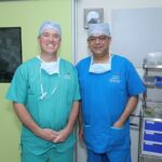
A 32-year-old woman successfully treated at AIMS Hospital, Dombivli, after developing serious complications related to a Ventriculoperitoneal (VP) shunt implanted at the age of eight following brain tumour surgery. The multidisciplinary team comprised Dr Ravi Sangle (Neurosurgeon), Dr Shahid Parvez (General and Laparoscopic Surgeon), Dr Rahul Jhankar (Neuro Physician), and Dr Deepak Chavan (Anaesthetist).
She presented with abdominal pain and fever for seven days and was found to have a CSF collection around the abdominal end of the shunt, along with redness, tenderness and a collection along the shunt pathway near the scalp. After the shunt was removed and cultures showed no infection, doctors identified a blockage in the normal flow of cerebrospinal fluid (CSF) and performed Endoscopic Third Ventriculostomy (ETV), helping her avoid another permanent shunt.
Sonalika Das, 32, a resident of Dombivli, working as Interior Designer had been leading a normal life for nearly 24 years after undergoing surgery for a brain tumour at the age of 8 years. A right-sided VP shunt was placed during her childhood treatment to drain excess CSF from the brain into the abdomen. She remained well for years until August 2026, when she developed abdominal pain and fever that continued for seven days. She visited AIMS Hospital on 3rd August 2026, where doctors found complications involving the long-standing shunt.
An ultrasound of the abdomen showed a lump in the pelvis surrounding the tip of the shunt. Examination by the neurosurgical team showed redness and tenderness along the shunt pathway, with a collection under the skin at the point where the shunt entered the scalp. She was started on antibiotics, and the team waited until her fever settled and blood counts stabilized before proceeding with surgery. The shunt was then removed with the help of diagnostic laparoscopy. During the procedure, doctors found that it was firmly attached to the surrounding tissues. Bowel loops were also stuck together around a collection of CSF in the abdomen. Samples of the CSF and the shunt tips were sent for culture, and the results were negative.
Dr Ravi Sangle , Neurosurgeon at AIMS Hospital, said, “VP shunt can work well for many years, but complications can sometimes develop much later due to blockage, infection or problems around the shunt. This is one of the uncommon but serious complications. In Sonalika’s case, the abdominal complication required us to remove the old shunt and first ensure that there was no ongoing infection.
In Sonalika’s case, this was considered because she had already experienced a significant complication with her previous shunt and had aqueductal stenosis; it may have happened as a foreign body inflammatory reaction towards shunt. The procedure was completed in 40 mins she was monitored for 4 days and was discharged on 5th September 2026 in stable condition. Not treating her at the right time could have led to further complications like Hydrocephalus, seizures, coma, if shunt inserted again then blockages and infection due to shunt.
Sonalika is doing perfectly well now after surgery and have resumed her routine life and work. “The entire experience was frightening for our family, especially because she had been doing well for so many years. We are thankful to the doctors and the entire AIMS Hospital team for giving her a chance to live without depending on a shunt again,” said the patient’s family.
The case highlights the need for timely evaluation of people with long-standing VP shunts when they develop new symptoms such as persistent headache, vomiting, fever, abdominal pain or redness and swelling along the shunt pathway. In selected patients with obstructive hydrocephalus, ETV can provide an alternative way to restore CSF flow and avoid placement of another permanent shunt.
Dr. Shahid Parvez, General Surgeon at AIMS Hospital said, the removal of a long-standing VP shunt can be challenging when it becomes firmly attached to surrounding tissues. Careful surgical intervention is essential to manage such complications safely and ensure the best possible outcome for the patient.”